An interactive tool meets students where they are, turning complex anatomy into something they can see, feel and explore.
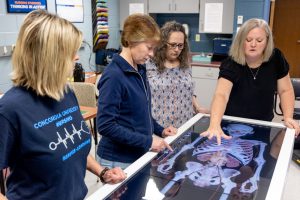
At first glance, it looks like an oversized touchscreen. But that impression doesn’t hold for long. Once it’s on and someone begins to move through it, it becomes something more.
The human body comes into view. Systems take shape on the screen, each one connected, each one able to be explored in more detail. With a swipe, a heart begins to beat. With another, it changes—no longer healthy, but struggling.
For students gathered around the table, the human body is no longer just something to study. It’s something to work through. Here, it even has a name.
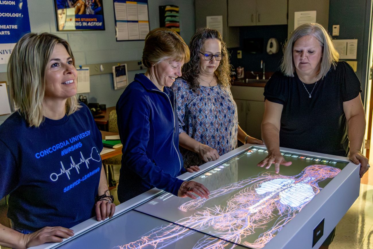
The first, “Hans,” is named after the first cadaver created by the Anatomage company. The second, “Cassie,” was donated by Rev. Dr. John Buuck, former Concordia president, and named in honor of his granddaughter, a Concordia nursing graduate.
For years, learning anatomy meant diagrams, models and, when possible, cadaver labs. Those methods still matter. But the Anatomage Table adds something different—something immediate.
“It’s one thing to talk about how the heart works,” said Tina Gaffney, MSN, RN, Simulation and VR Center director and a 2022 CUW graduate. “It’s another to stand here and actually see it happening.”
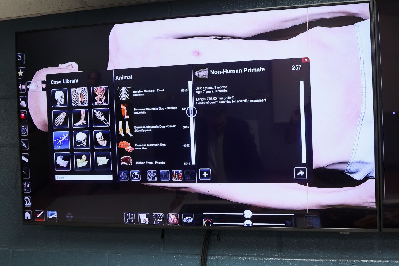
Instead of imagining how systems connect, students can follow them in real time. A normal organ can sit beside one affected by disease. A single screen can split into multiple views, allowing comparisons that would be difficult to recreate any other way.
And it’s not just about observation. Students can trace pathways, isolate structures and even simulate procedures. In one setting, they might walk through the stages of a colonoscopy. In another, they can watch a birth unfold from an internal perspective—something that shifts how they understand what’s happening beyond the surface.
How it shows up in the classroom
In Concordia’s nursing program, the table is already finding its place alongside simulation. Students might begin with a patient scenario—responding, making decisions and working through what comes next. Then, almost immediately, they move to the table.
“We can take what they just experienced and show them what’s happening inside the body,” Gaffney said. “That connection makes a difference.”
During cardiac simulations, students don’t just talk about heart function. They see it. They can watch how electrical activity changes the way the heart pumps, and how that shift affects the rest of the body. It’s a step that brings the classroom closer to real practice.
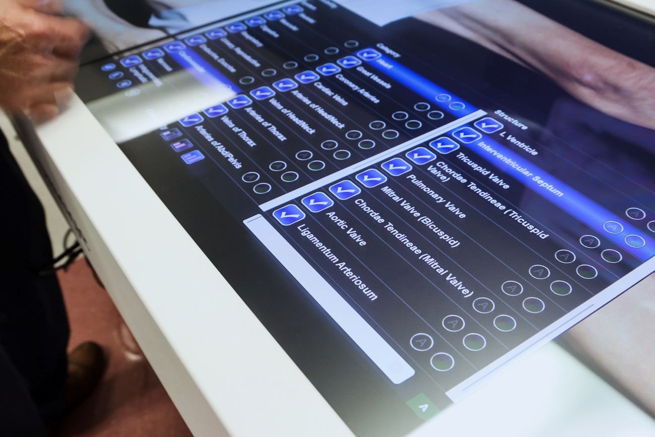
There’s also a certain ease students have when they approach the table. It doesn’t feel foreign. They move through it the way they’ve learned to move through most things—by touching, exploring and testing.
“Students aren’t just standing back and listening,” Gaffney said. “They’re putting their hands on it and figuring it out.”
That matters, especially as Concordia continues to move toward competency-based education. Understanding isn’t just about recalling information. It’s about being able to show it, apply it and explain it. Here, students can do exactly that.
Across programs
While nursing has been one of the first areas to use the table, it’s not the only one. Faculty across campus are beginning to see where it fits into their own teaching.
In occupational therapy, movement becomes easier to visualize. Students can manipulate joints, follow muscles through motion, and begin to see how structure supports everyday function.
“The possibilities for exploring anatomical structures with the Anatomage Table are truly endless,” said Jessica Mischler (’14), a certified hand therapist and adjunct faculty member in Concordia’s Occupational Therapy program.
She notes how easily users can shift between supine and prone positions, allowing students to view the body from multiple perspectives in just moments. Unlike traditional dissection, the table enables virtual tissue retraction without permanent removal, giving students the chance to revisit and study structures as often as needed. For Mischler, that flexibility matters. It creates a hands-on learning experience that supports repetition, curiosity, and deeper understanding—something not possible with a live donor alone.
That flexibility also removes some of the common barriers students face in lab settings.
“The ease of access to the human body with the Anatomage Table is truly irreplaceable,” Mischler said. “There’s no need for setup, protective equipment or cleanup. By removing these logistical barriers, students can focus on mastering complex anatomical information.”
In biology, the level of detail offers something beyond static images. Students can work through the various systems of the body, focusing on how structures connect and function together.
In her anatomy and physiology course this past spring, Dr. Sarah Lovern, professor of biology, introduced the Anatomage Table as part of the lab experience. Students used it to identify bones and anatomical regions across different cadavers, gaining a clearer sense of size and spatial relationships.
“They liked seeing the bones in real sizes on the specimens,” Lovern said. “They also practiced learning all of the anatomical terms for the regions of the body.”
Students responded positively to the hands-on approach, especially as they interacted with and manipulated the images on screen. The table gave them a new way to engage material often limited to diagrams or models.
Mischler noted that the technology can also make anatomy more approachable for students who may feel uncertain entering a traditional lab environment.
“The Anatomage Table removes barriers that might otherwise intimidate students or interfere with learning the body’s complex functions,” she said. “It creates a more accessible environment and helps students engage more fully with the material.”
Looking ahead, Lovern plans to expand how the table is used in the classroom. Students will work through case studies that explore how body systems interact, with added focus on the circulatory system, as well as the endocrine and respiratory systems.
“We will continue to build on these skills next semester,” she said.
As more faculty across programs integrate the table into their teaching, its place in the curriculum will continue to grow.
It starts with a real person
The Anatomage Table isn’t replacing what’s already in place. Students will still need to read and take notes. They will still practice in clinical settings. Those pieces remain essential.
What this adds is another way in—another way to understand something that can be difficult to picture.
“There’s still a place for all of it,” Gaffney said. “This just helps bring it together.”
While the images on the table appear digital, each one begins with something deeply human. Individuals chose to donate their bodies to science, and that reality remains central to the learning experience.
As the table becomes more integrated into coursework, faculty are also thinking through how to approach it with care—how to remind students that what they are seeing represents real people.
It’s a perspective that aligns naturally with Concordia’s mission of developing students in mind, body and spirit.
The stories it holds
Right now, the table is still new. Faculty training is ongoing. Departments are exploring how it fits. This upcoming fall, students will begin working with it more consistently. There’s still more to learn about how it will shape the classroom.
But standing around it, watching students interact with something that once lived only in textbooks, it’s clear that something has already shifted. Today’s students are multimodal learners, engaging with material in ways that invite deeper understanding and lasting connection.
“It gives them a clearer picture,” Gaffney said. “And once they can see it, they start to understand it differently.”
And that understanding matters. It carries forward into labs, into clinical settings and eventually into the lives of the patients and communities they will serve.
At Concordia, tools like the Anatomage Table do more than introduce new technology into the classroom. They prepare students with the knowledge, care and purpose needed for the work they are called to do.
How it is used
At Concordia, the Anatomage Table is beginning to support learning across programs in ways that extend beyond a traditional lab, with additional applications continuing to take shape.
Students and faculty can use the table to:
- Explore full-body anatomy in 3D, moving through systems and structures in real time
- Perform virtual dissections and examine organs from multiple angles
- Identify anatomical regions and structures on life-sized cadaver images
- Compare healthy and diseased anatomy side by side
- Simulate clinical procedures, including colonoscopies and endoscopies
- Visualize processes such as childbirth from an internal perspective
- Pair anatomy with simulation scenarios, such as CPR and cardiac response
- Analyze movement of joints and muscles for occupational and physical therapy
- Study brain structure and function, including the effects of injury or neurological conditions
- Examine facial muscles and jaw movement for speech-related applications
- View histology slides, including cancerous and damaged tissue
- Complete quizzes and hands-on assessments by identifying structures directly on the table
- Explore how body systems connect and interact through case-based learning
- Support patient education by visualizing procedures and conditions
The table is updated annually and supports both in-person and remote instruction, giving faculty flexibility in how they teach and how students engage.
Want in?
Concordia University Wisconsin is a Lutheran higher education community committed to helping students develop in mind, body and spirit for service to Christ in the Church and the world.